Rappoport's environment is an enrichment medium and a differential diagnostic medium. In her compound includes: bile broth 10%, 2% glucose, indicator (acid fuchsin, discolored with alkali; colorless in an alkaline environment, red in an acidic environment). Purpose: for the accumulation of typhoid-paratyphoid bacteria when inoculating the patient’s blood, as well as for the approximate differentiation of typhoid-paratyphoid bacteria. Principle d: with the growth of typhoid-paratyphoid bacteria due to glucose fermentation, diffuse turbidity and redness of the medium occurs. For paratyphoid bacteria, in contrast to typhoid bacteria, the presence of gas in the float.
Endo medium – differential diagnostic. Compound: MPA, lactose, indicator – basic fuchsin, decolorized with sodium sulfite. Purpose: for sieving the test material in order to obtain isolated colonies. Principle d-i is based on the ability of some microorganisms to decompose or not decompose lactose (salmonella does not decompose and forms colorless colonies).
Wednesday Levin is weakly selective and differential diagnostic. In her composition MPA, tactose, indicators: eosin and methylene blue, potassium hydrogen phosphate. For purpose and principle, see Endo environment.
Russell medium Compound: MPA, lactose 1%, glucose 0.1%, indicator (aqueous blue and rosolic acid; pink in an alkaline environment, blue in an acidic environment). Purpose: for screening out “suspicious” colonies in order to isolate pure cultures and determine their enzymatic activity towards glucose and lactose. Principle d:
Kligler's medium – combined, differential diagnostic. Compound: MPA, lactose 1%, glucose 0.1%, sodium thiosulfate, iron II sulfate, indicator - phenol red (in an alkaline environment - red, in an acidic environment - yellow). Purpose: for screening out “suspicious” colonies in order to identify pure cultures and determine their enzymatic activity towards glucose, lactose and H2S formation. Principle d: enterobacteria, which have the enzyme thiosulfate reductase, reduce thiosulfate to sulfite, and hydrogen sulfide is released, which reacts with iron sulfate - resulting in the formation of black iron sulfide.
Serological identification of an isolated pure culture using adsorbed monoreceptor O- and H-agglutinating Salmonella sera. Serological identification is carried out in an agglutination reaction on a stack with agglutinating adsorbed Salmonella O- and H-sera; these sera may 2 types: monoreceptor and polyvalent, they are obtained from specific immune sera by the method of antibody adsorption according to Castellani.
Serological identification proceeds sequentially in 3 stages:
1) Establishment of the culture’s belonging to serogroups A, B, C, D, E, of the Salmonella genus. In this case, a polyvalent O-serum is used containing antibodies against antigen receptors 2, 3, 4, 7, 9, 10. Monoreceptor O-serums are used against rare serogroups. When diagnosing paratyphoid A and B and typhoid fever, you can use a mixture of O-sera containing antibodies to receptors 2, 4, 9.
2) If the result is positive, to determine whether the test culture belongs to a specific serogroup, an agglutination test is performed separately with each monoreceptor O-serum included in the polyvalent serum: receptor 2 belongs to serogroup A, receptor 4 to serogroup B, receptor 7 to to serogroup C, etc.
3) After determining the serogroup, we determine its species using the agglutination method with adsorbed monoreceptor H-sera against the H-antigens of the 1st phase of salmonella that are part of this serogroup. Then agglutination is carried out with adsorbed H-sera against H-antigens of the 2nd phase and the antigenic formula of the test culture is finally established.
Bacterial carriage in typhoid fever. What serological reactions can confirm chronic bacterial carriage? Diagnosticums used for this purpose. The only evidence of bacterial carriage is the isolation from the carrier of cultures S. typhi, S. paratyphi A, S. paratyphi B, using auxiliary methods: serological reaction and allergic skin test with Vi-typhin (it contains Vi-antigen, which, when interacting with Vi-antibodies gives a local allergic reaction in the form of redness and swelling for 20-30 minutes). A positive reaction with Vi-typhine indicates that the organism contains Vi-antibodies and possibly S. typhi.
Chr. bacterial carriage is confirmed by RNGA. In this case, Vi-diagnosticum is used, because during the formation of typhoid bacteria carriage, the manifestation of Vi-antibodies is typical; diagnostic titer of the solution is 1:40.
Methods of infection with typhoid fever. Source of infection. Infection with typhoid fever occurs only through the oral route. The source of infection is sick people and bacteria carriers (they pose a danger to others for several years after the illness).
Composition (in terms of 1 liter of prepared medium):
Enzymatic peptone, dry - 10.0 g.
Autolyzed yeast extract clarified - 1.5 g.
Lactose - 10.0 g.
Sodium phosphate disubstituted - 2.0 g.
Microbiological agar - 13.0 g.
Sodium chloride - 3.0 g.
Eosin sodium, indicator - 0.4 g.
Methylene blue, indicator - 0.075 g.
A dense nutrient medium for the isolation and differentiation of enterobacteria from the test material based on lactose fermentation.
Mix the dry medium in an amount of 40 g in 1 liter of purified water, bring to a boil and boil until the agar is completely melted (2-3 minutes), filter through a cotton-gauze filter, pour into sterile bottles and sterilize by autoclaving at a temperature of 112 ° C for 20 min. Cool the sterile medium to a temperature of 45-48oC and pour into sterile Petri dishes; After the agar has hardened, dry the dishes at a temperature of 37°C for 40-60 minutes. The color of the finished medium should be reddish brick.
Before use, the prepared medium can be stored in a dark place for no more than 7 days at a temperature of 2-8°C.
Incubate the inoculations of the test samples for 18-48 hours at a temperature of 37°C. Visual growth control is based on the presence or absence and appearance of colonies. Lactose-positive microorganisms form opaque colonies from light lilac to violet with or without a metallic sheen. Lactose-negative microorganisms form transparent or translucent colorless colonies (they can form pale pink colonies).
Disposal of dry media with expired shelf life and used ready-made culture media - in accordance with the requirements of SanPiN 2.1.7.728-99 Rules for the collection, storage and disposal of waste from medical institutions.
Storage- in the manufacturer's packaging in a dry place, protected from light, at a temperature from 2°C to 25°C. Freezing is not allowed.
Transportation - at temperatures from 2°C to 25°C. Freezing is not allowed.
LEVINA WEDNESDAY(M. Levine, American bacteriologist, born in 1889; syn. eosin methylene blue agar) - colored selective nutrient medium for differentiation of bacteria of the family. Enterobacteriaceae is used in the laboratory diagnosis of dysentery, typhoid fever, salmonellosis and other intestinal infections. Eosin and methylene blue agar was first proposed in 1916 by J. E. Holt-Harris and O. Teague; later, in 1918, the composition of the medium was modified by Lewin.
L.S. along with other solid colored differential diagnostic nutrient media for enterobacteria, has found wide use in the laboratory. practice. With its help, it is possible to detect pathogenic lactose-negative microflora in a relatively large percentage of cases. Ready HP quite stable, does not change its properties in the light or when stored for several days; its manufacture does not require precise determination of the concentration of hydrogen ions (pH). Included in the composition of HP. organic dyes selectively inhibit the growth of gram-positive bacteria, due to which it is both an enrichment medium and gives relatively better results when directly inoculating the test material, even if it contains a relatively small number of pathogenic bacteria.
Depending on the quality of individual ingredients (peptone and especially dyes), the resulting HP. the results may vary somewhat, therefore, when preparing each new series of media, it is advisable to use materials of the same brand, which greatly facilitates the recognition of colonies.
Several recipes and methods for preparing HP are known. (Yu. A. Kozlov, G. Ya. Sinai, O. G. Birger, etc.). The most widely used method is one in which the basic medium, solutions of lactose and dyes are prepared separately, suitable for preservation for future use and mixed before use.
The basic medium consists of 10 g of bacteriological peptone, 15 g of agar-agar, 2 g of dipotassium phosphate (K2HPO4) and 1 liter of distilled water. It is sterilized in an autoclave at 1 atm for 15-20 minutes, the pH is not adjusted.
When preparing a differential medium, for every 100 ml of molten basic medium, with constant stirring, add the following solutions prepared in distilled water and pre-sterilized with flowing steam (fractionally, 3 days in a row for 15-20 minutes each) in the given sequence 5 ml of 20% solution ra of medical lactose, 2 ml of 2% alkaline eosin solution (bacteriological), 1.5 ml of 0.5% methylene blue solution. The prepared medium is poured into Petri dishes, slightly dried and used as usual. The color of the medium is blue-violet.
Colonies of Escherichia coli on L. p. small, round, with a smooth shiny surface, dark blue (to black) color, sometimes with a metallic sheen. Young colonies are cloudy, opaque, and can only be colored in the center. Colonies of pathogens of dysentery, typhoid fever and paratyphoid fever are relatively smaller, round, shiny and transparent, usually completely colorless (young) or with a slight pinkish or bluish tint. Protea colonies do not produce creeping growth; they are small, isolated, and orange-yellow in color. The medium changes color only around Protea colonies.
Some authors recommend increasing the amount of 0.5% methylene blue solution to 2 ml per 100 ml of the basic medium or eliminating the phosphate buffer from its composition. You can prepare the medium not with peptone, but based on Hottinger’s digest, at pH 7.2-7.3 with the addition of the appropriate amount of agar and potassium phosphate. The use of meat water for preparing the medium is unacceptable; the differentiation of colonies in this case is significantly worse.
There is a known method for preparing dry HP, which is very stable in quality and especially convenient for long-term storage and under conditions of expeditionary work (N.V. Ploskirev).
Stable dry HP. is produced by industry, instructions for its manufacture and use are sent along with the medium. Most diagnostic laboratories use high-quality standard selective dry nutrient media of domestic production for the isolation and differentiation of pathogenic enterobacteria - L. s., Bacto-agar F, Ploskirev's medium (see Ploskirev's medium).
Bibliography: Guide to microbiological diagnosis of infectious diseases, ed. K. I. Matveeva, p. 110, M., 1973; Handbook of microbiological and virological research methods, ed. M. O. Birger, p. 58, M., 1973; Levine M. The effect of concentration of dyes on differentiation of enteric bacteria on eosin-methylene-blue agar, J. Bact., v. 45, p. 471, 1943.
G. P. Belikov.
Levina Wednesday Levina Wednesday
(lactose-eosin-methylene agar) is a differential-selective medium for the isolation of enterobacteria. Allows you to distinguish lactose-fermenters (form dark blue or black colonies) from non-lactose-fermenters (colonies the color of the medium). Proteus forms isolated orange colonies. The medium is prepared by adding to 100 ml of 1.5% MPA, pH 7.2 - 7.4, 2 ml of 0.5% aqueous solution of methylene blue, 1.5 ml of 2% eosin solution, 2 g of lactose and 0 .2 g of dibasic potassium phosphate, then pour it into Petri dishes. The medium is light purple in color. The industry produces a dry preparation, which is prepared according to the instructions on the label. Option HP -lactose bromide-mol medium - does not have inhibitory properties against Klebsiella scleroma.
(Source: Dictionary of Microbiology Terms)
See what “Levina Wednesday” is in other dictionaries:
- (M. Levine, born in 1889, American bacteriologist) a dense differential diagnostic nutrient medium for identifying pathogenic bacteria of the family that do not decompose lactose. Enterobacteriaceae (causative agents of typhoid fever, dysentery, etc.); consists of... ... Large medical dictionary
- (s) (s) an artificial substrate, which is a balanced mixture of nutrients in concentrations and combinations necessary for the growth and division of microorganisms or cells of higher organisms. Adams's nutrient medium see Adams... ... Medical encyclopedia
See Levin Wednesday... Large medical dictionary
See Levin Wednesday (Source: “Dictionary of Microbiology Terms”) ... Dictionary of microbiology
- (tetrabromofluorescein) dye (see Dyes) of red color and fluorochrome of greenish yellow color. Soluble in water and ethanol. Used for dyeing cytoplasm and as a composition of Romanovsky Giemsa paint, and also as an inhibitor of certain bacteria in... ... Dictionary of microbiology
This term has other meanings, see Children's music school No. 1. Children's music school No. 1 in Penza ... Wikipedia
Culturological and social psychological theories that describe and explain human beings. behavior and social reality through interactions between people. Elements T.v. contained in the sociology of Simmel, the phenomenology of Husserl and... ... Encyclopedia of Cultural Studies
- (co-infections) diseases caused by bacteria of the genus Escherichia (see). Obligate enteropathogenic variants of Escherichia coli cause the specific disease colienteritis (see). Wedge, picture of E., caused by opportunistic intestinal... ... Dictionary of microbiology
General and scientific term meaning: 1) human. the individual as a subject of relations and consciousness. activity (person, in the broad sense of the word) or 2) a stable system of socially significant traits that characterize the individual as a member of this or... Philosophical Encyclopedia
T.P. was created by Kurt Lewin, who believed that to understand behavior it is necessary to take into account the entire situation, i.e. the gestalt situation. Levin extended his ideas to new areas, taking mat as a model of thinking. construction... ... Psychological Encyclopedia
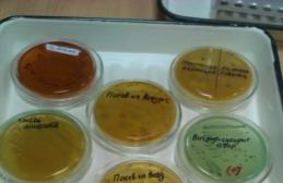
Practice
Dense media


Bismuth sulfite agar
______________________________________________


Paratyphoid A Salmonellosis
The prepared medium is opaque and pea-green in color. Strictly selective medium for isolating pure Salmonella cultures. Salmonella typhi, Salmonella enteritidis And Salmonella typhimurium usually form on this medium black colonies with a metallic sheen, surrounded by a zone of blackening as a result of the production of hydrogen sulfide and the reduction of sulfite to iron sulfide, which has a black color. Salmonella paratyphi A forms light green colonies.
Sample answer: Small colonies were found on the dense medium “Bismuth-sulfite agar”. Smooth with an even edge, dark and opaque with a metallic sheen.
______________________________________________
Wednesday Levin


Sample answer: On this medium, Levin's medium, small smooth colonies with a smooth edge of a blue-violet color with a metallic sheen were found, with fermentation of lactose to acid. The indicator is methylene blue. Preliminary diagnosis - colienteritis caused by Escherichia coli - Escherichia coli.


Sample answer: This medium is Levin's medium, made for the purpose of obtaining isolated colonies. Small, smooth, transparent, uncolored colonies were found on the medium, and lactose fermentation was absent. Pre-diagnosis – dysentery caused by possible pathogens:
Serogroup A: Shigella dysenteriae(10 serovars)
Serogroup B: S. flexneri(6 serovars and subtypes)
Serogroup C: S. boydii(15 serovars)
Serogroup D: S. sonnei(1 serovars)
______________________________________________
Endo medium
______________________________________________


Sample answer: This medium is the Endo medium. Small, colorless, transparent, smooth colonies with a smooth edge were found on it. There is no decomposition of lactose to acid, as indicated by the color of the medium. The Andrade indicator is used. The presumed diagnosis is typhoid fever caused by Salmonella Typhi.


Sample answer: This medium is used for the accumulation of isolated colonies. Wednesday – Endo. Found small, smooth with a smooth edge, red with a metallic sheen due to the decomposition of lactose to acid using the Andrade indicator. Preliminary diagnosis - colienteritis caused by Escherichia coli - Escherichia coli.
______________________________________________
Methods for determining sensitivity to antibiotics
______________________________________________

Sample answer: The Petri dish introduces a method for determining antibiotic sensitivity - the paper disc method, in which a bacterial suspension is applied to the surface of the agar in the Petri dish and then discs containing a certain amount of antibiotic are placed. Diffusion of the antibiotic into the agar leads to the formation of a zone of suppression of the growth of microorganisms around the disks. After incubating the dishes in a thermostat at a temperature of 35 o -37 o C overnight, the result is taken into account by measuring the diameter of the zone around the disk in millimeters.
The isolated culture is highly sensitive to monomycin, the diameter to growth retardation is 30 mm, neomycin - 26 mm, kanamycin - 20, and no chloramphenicol at all. Characteristic of E. coli - Escherichia coli.
______________________________________________
Seeding from the air
______________________________________________


Sample answer: This plate agar was inoculated from air using the sedimentation method. An indication of indoor air pollution is the total number of bacteria per 1 cubic meter. The number of bacteria is determined in 10 minutes. To do this, the Petri dish with the plate MPA is left open in the air for 10 minutes, then incubated in a thermostat at a temperature of 37 degrees for 24 hours. The results are calculated by the total number of colonies. The norm in the operating room before surgery is 3-5 colonies. After – 15. There is also an aspiration method, where the air is assessed with a special Krotovy device.
______________________________________________
Mixture of microorganisms
______________________________________________

Color range
______________________________________________
Dysentery
______________________________________________


MPB (for determining proteolytic properties): H2S-; Indole-;
Mannitol: Acid+; Gas-;
Maltose: Acid+; Gas-;
Peshkov's medium: growth according to the prick, the color has changed to red, which indicates the fermentation of mannitol to acid. Indicator – Andrade;
______________________________________________
Escherichia coli
______________________________________________


MPB (for determining proteolytic properties): H2S+ (blackened); Indole+(blushed);
Glucose: Acid+; Gas+;
Lactose: Acid+; Gas+;
Maltose: Acid+; Gas+;
Sucrose: Acid-; Gas-;
Peshkov's medium: turbidity of the entire column (the culture is mobile), the color has changed to red, which indicates the fermentation of mannitol to acid and the presence of gas bubbles. Indicator – Andrade;
Ressel's medium (Indicator - bromothymol blue): The bevel and column are yellow, and in the thickness there is gas, which indicates the decomposition of lactose and fructose to gas and acid.
Typhoid fever
______________________________________________


Hiss medium (Indicator - water-rosol):
Glucose: Acid+; Gas-;
Maltose: Acid+; Gas-;
Mannitol: Acid+; Gas-;
Lactose: Acid-; Gas-;
Sucrose: Acid-; Gas-;
Peshkov's medium: turbidity of the entire column (the culture is mobile), the color has changed to red, which indicates the fermentation of mannitol to acid. There is no gas. Indicator – Andrade;
______________________________________________
Salmonellosis
______________________________________________


MPB (for determining proteolytic properties): H2S+ (blackened); Indole-;
Hiss medium (Indicator - water-rosol):
Maltose: Acid+; Gas-;
Sucrose: Acid-; Gas-;
Peshkov's medium: turbidity of the entire column (the culture is mobile), the color has changed to red, which indicates the fermentation of mannitol to acid and gas. Indicator – Andrade;
Ressel's medium (Indicator - bromothymol blue): The slope has an unchanged color, and the column is yellow. There is gas in the thickness, which indicates the decomposition of fructose into gas and acid; lactose is unfermented.
______________________________________________
Gas gangrene
______________________________________________


Kitta-Tarozzi medium: liquid medium for the cultivation of anaerobes. To prepare it, liver (meat) cut into small pieces is poured with three times the amount of MPB, boiled for 30 minutes, and filtered. Dried pieces of liver are placed (for oxygen adsorption) into test tubes and filled with broth. Sterilize at 120°C for 30 minutes. Before use, warm up and fill with sterile neutral paraffin oil.
Growth in the form of turbidity was detected on this medium. There is also gas formation.
______________________________________________
Botulism
______________________________________________


Weinberg's medium: a semi-liquid nutrient medium used for the cultivation of pathogenic anaerobes, which is a meat-peptone broth containing 1% agar and 0.2% glucose;
Small, disc-shaped, fluffy colonies were found in the sugar agar column. The medium is ruptured due to increased gas production during glucose fermentation.
They make a cut. Using a Pasteur pipette or needle, take part of the colonies and place them in Kitta-Tarozzi medium for accumulation.
______________________________________________
Agglutination reaction (expanded)
______________________________________________


NB!: Friends, do not forget that you need to read reactions from the end - checking serum and antigen control. Afterwards you will most likely be asked for a definition agglutinating serum titer (the maximum dilution of serum at which agglutination with the corresponding microorganism occurs) And diagnostic serum titer (titer of antibodies to a specific pathogen in the blood serum, detected in patients and not observed in healthy people).
Sample answer: This reaction is a detailed agglutination reaction. The serum control appears as a clear liquid and is negative. Antigen control shows turbidity, also negative. We look at the dilution of 1/100 - transparent with an umbrella at the bottom, when shaken - flakes, which means the reaction is positive. And so on up to 1/6400. We look at the dilution of 1/12800, there is noticeable turbidity - the reaction is negative.
Consequently, the agglutination reaction is positive up to a dilution of 1/6400, with negative controls, which means the titer of the test serum is 1/6400.
An agglutination reaction is the gluing of corpuscles (bacteria, red blood cells, etc.) by antibodies in the presence of electrolytes - sodium chloride.
______________________________________________
Compliment binding reaction
______________________________________________


RSC is based on the activation of complement by the antigen-antibody complex.
It takes place in two stages:
Incubation of the mixture: antigen+antibody+complement
Indicator: detection of one complement by adding a hemolytic system (sheep red blood cells + hemolytic serum in triple titer)
Sample answer: CSCs were recorded with the sera of patients 1 and 2 to identify antibodies to the corresponding antigen in them. The reaction with the serum of patient 2 is negative, since in the first stage the antibody and antigen did not match each other and did not bind, which means they did not bind complement. Then free complement combines with the hemolytic system and lysis occurs. Delayed hemolysis was detected in the serum of patient 1, which means that an “antibody+antigen+complement” complex has formed. Complement is bound and does not attach to the hemolytic system. There is no hemolysis. The diagnosis is positive.
______________________________________________
Passive (indirect) hemagglutination reaction
______________________________________________

IN RNGA detect serum antibodies using antigenic erythrocyte diagnosticum, which are red blood cells with antigens adsorbed on them.
RPGA placed in plastic tablets or in test tubes with dilutions of the patient’s blood serum, to which an erythrocyte diagnosticum is added.
Red blood cells (or latex particles) with antigens adsorbed on them interact with the corresponding antibodies in the blood serum, which causes the red blood cells to stick together and fall out to the bottom of the test tube or cell in the form of a scalloped sediment. In a negative reaction, red blood cells settle in the form of a button.